Address
10 place de la Joliette
Atrium 10.4 – Etage 6
13002 Marseille, France
With the advanced capabilities of our BondXplorer™ microscope and InSplorer™ endoscope, we are equipped to offer a diverse range of imaging modalities for plant studies. One of the standout features is our ability to perform nonlinear fluorescence imaging, which provides exceptional detail and insight into plant structures and processes.
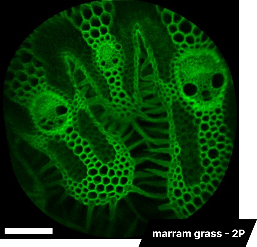
Marram grass 2P imaging
InSplorer endoscope
The image on the left showcases an example of endoscopic 2-photon (2P) fluorescence imaging conducted on a marram grass sample. The lignified cell walls, which have been labeled, emit a strong fluorescence signal, making them highly visible. On the other hand, the cellulose is less visible but still discernible. The scale bar in the image represents 100 µm. This demonstrates the power and precision of our imaging technology in revealing intricate details of plant structures.
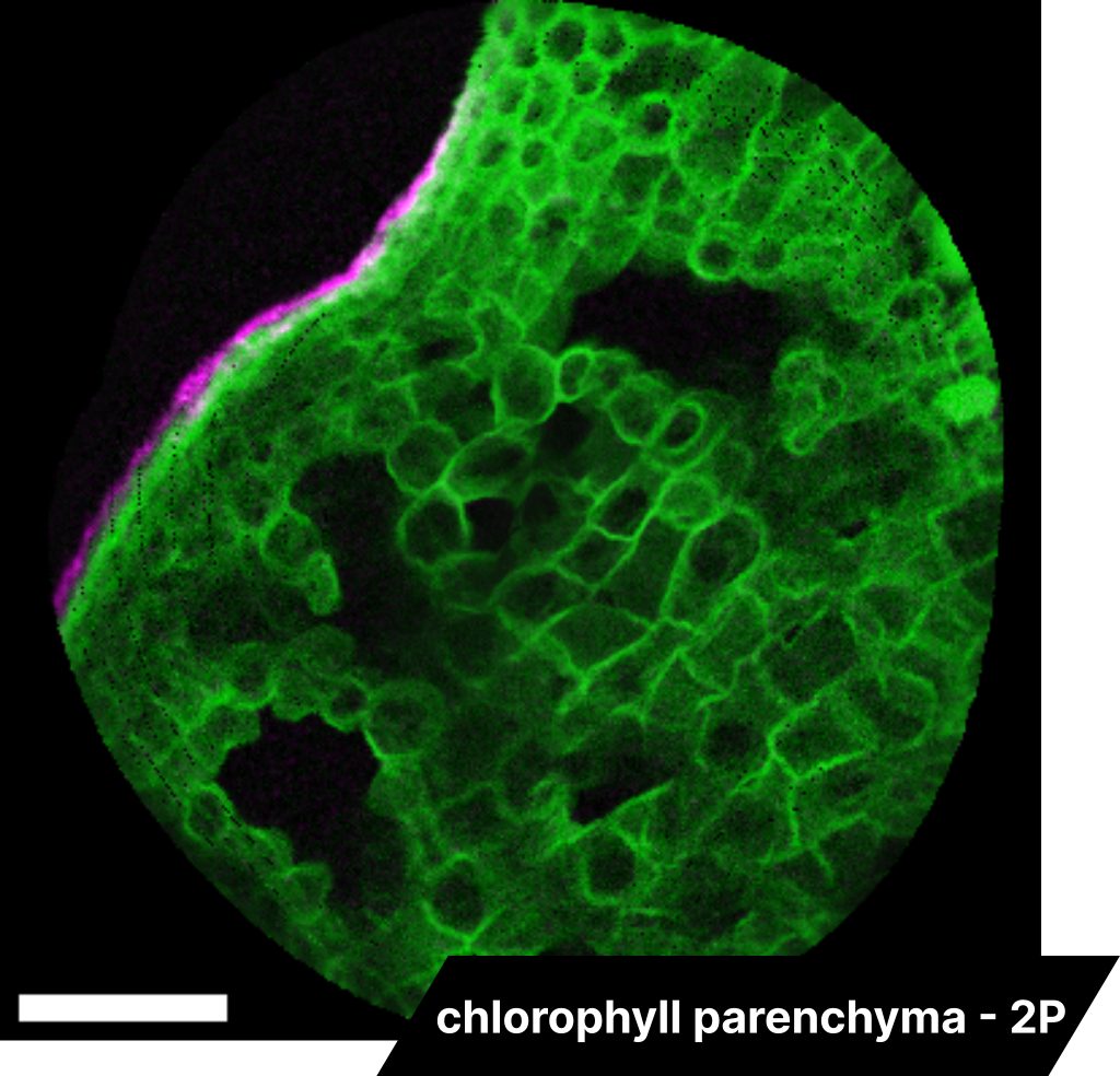
Parenchyma 2P imaging
InSplorer endoscope
Leveraging a single excitation source at 860 nm, we carry out endoscopic 2-photon imaging on chlorophyll parenchyma. By employing a collection filter set at 520 nm, we can detect the autofluorescence of lignin, which is represented in magenta. Additionally, stained cells are visible in green, using a filter at 630 nm. The scale bar in the image corresponds to 100 µm.
Oak leaf SRS and 2P imaging
BonXplorer microscope
The dual color z-stack image of an oak leaf, CH3-bond SRS signal in cyan and two-photon fluorescence in magenta, was obtained from the scanning of the leaf in depth with the BondXplorer™ microscope, thereby revealing stomatas and cells. The scale bar in the image represents 43 µm.
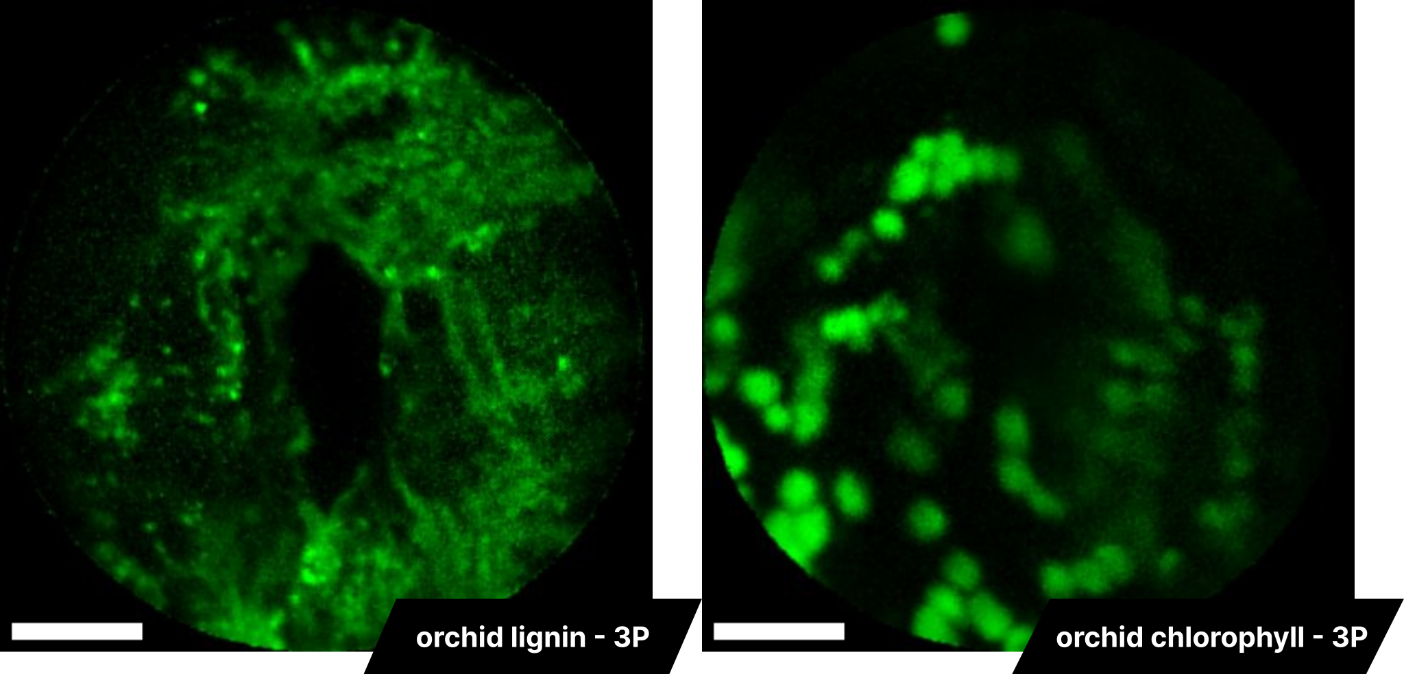
Orchid 3P imaging
InSplorer endoscope
By harnessing the capabilities of the InSplorer™ endoscope, we are able to conduct 3-photon (3P) fluorescence imaging. The image provided is an example of 3P imaging performed on an orchid sample. Utilizing a single excitation source at 1300 nm, we can selectively image either lignin or chlorophyll by employing a collection filter at 520 nm or 630 nm respectively. The scale bar in the image represents 20 µm.